


Proteinuria in glomerular disease
PROTEINURIA IS A KEY FEATURE OF GLOMERULAR DISEASE1-4
Risk factor:
Proteinuria is a major RISK
FACTOR for glomerular disease, if left unchecked it can lead to end-stage kidney disease (ESKD), dialysis, and even death2–6
Predictor:
Proteinuria is the single strongest modifiable PREDICTOR of the rate of kidney function decline in glomerular diseases such as IgA nephropathy (IgAN)1,2,5,6
Glomerular diseases:
Glomerular diseases such as IgAN are characterized by high levels of proteinuria6

In IgAN:
Each incremental gram per day above 1g results in a 10- to 25-fold faster rate of decline in kidney function and survival6
View IgAN data

In patients with biopsy proven IgAN (n=542), proteinuria levels over time was the strongest predictor of the rate of kidney function decline6*
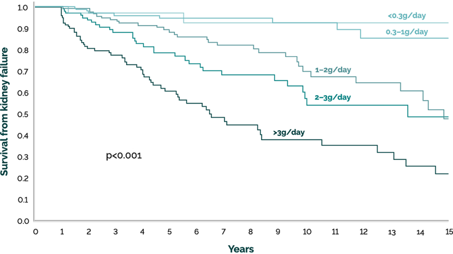
Adapted from Reich HN, et al. 20076
Each incremental gram per day above 1 results in a 10- to 25-fold faster rate of decline in kidney function and survival.6
*Data from a cohort of 542 patients with primary IgAN and were followed longitudinally in the Toronto Glomerulonephritis Registry. Time-average proteinuria was the most important predictor of renal survival (multivariate hazard 1.57; 95% confidence interval 1.39 to 1.77; P<0.01)6
Proteinuria is a key factor in the progression of IgAN
Proteinuria contributes to disease progression1,4,5,7
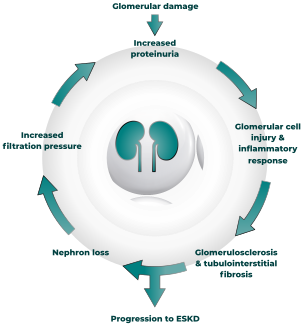
Proteinuria is primarily associated with a progressive loss of kidney function leading to kidney failure.5,9 The pathophysiologic link between proteinuria and progressively declining kidney function is complex, however there is evidence to suggest that tubular epithelial cells play a central role4
This effect of proteinxuria in disease progression occurs through various processes:1,4,5,7,8

Induction of tubular chemokine expression and complexment activation, leading to inflammatory interstitial cell infiltration and sustained fibrogenesis1,4,5,7,8

Activated angiotensin II (ANG II) produces vasoactive, inflammatory, and profibrotic cytokines, including the vasoconstrictor, endothelin-1 (ET-1)4,7,8

Disease progression is accelerated by a cycle of inflammation and activated tubular response, leading to tubular damage/apoptosis, podocyte damage, and ultimately interstitial fibrosis4,8
Proteinuria is a key marker of prognosis and disease severity in IgAN2,5,9,10
Proteinuria and reduced estimated glomerular filtration rate (eGFR) are joint contributors to glomerular disease progression2,5
Kidney function in IgAN
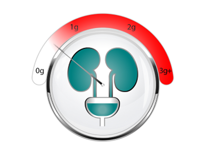
High proteinuria and declining eGFR are associated with an increased risk of disease progression in IgAN2,3
Slower decline in kidney function
Higher proteinuria levels are associated with a faster decline in eGFR;3,5 conversely, patients within normal proteinuria levels show a slower decline in eGFR2
Increased proteinuria was a stronger predictor of disease progression than a reduced eGFR2
Target proteinuria to slow progression to kidney failure
Reducing proteinuria is associated with improved patient outcomes1,4-6,10-16
Proteinuria is a modifiable risk factor: the more it is reduced, the greater the protective effect against decline in kidney function to ESKD1,4–6,10–16
In patients with IgAN (N=81):
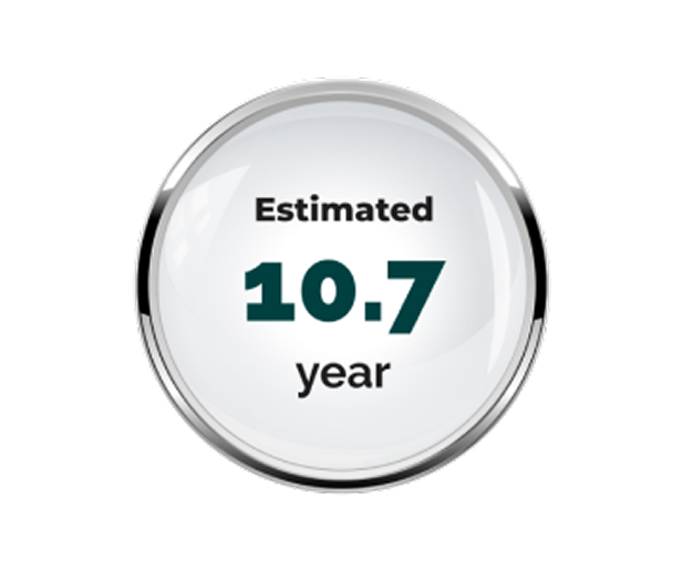
delay in the median time to kidney failure14
In IgAN, 30% reduction in proteinuria is estimated to reduce risk for ESKD by 50%, which is consequently estimated to increase the median time to ESKD. The median time to ESKD or eGFR <15mL/min/1.73 m2 was extended in this model from 12.4 years to 23.1 years14
View IgAN data
In IgAN, 30% reduction in proteinuria is estimated to reduce risk for ESKD by 50%, which is consequently estimated to increase the median time to ESKD14†
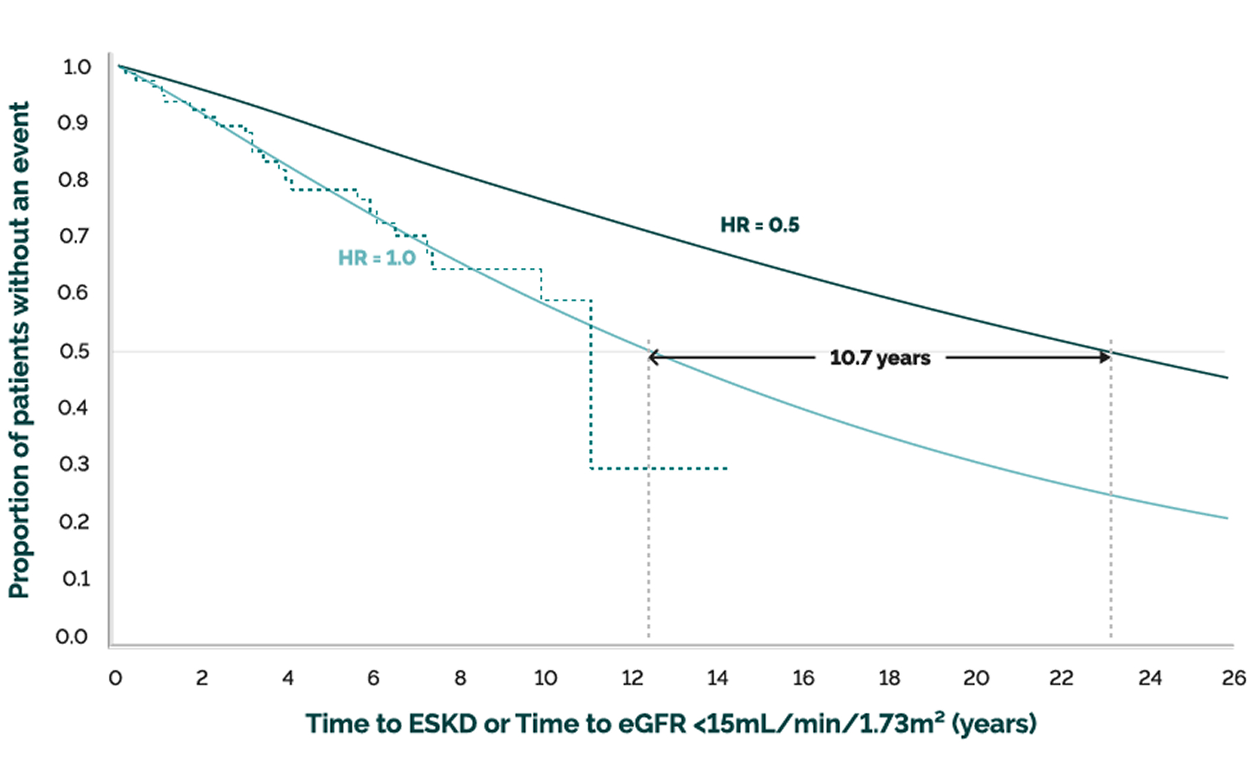
Adapted from Carroll K, et al. 202014
Patient inclusion criteria: IgAN patients with proteinuria value ≥1.0g/day or UP/C ≥1.0g/g and eGFR ≥30mL/min/1.73 m2 at the initiation of RAS blockade.14
†Dataset from Leicester University Hospitals record of IgAN patients (n=81). Time to ESKD was estimated via a Kaplan Meier plot & Weibull fit analysis. HR=1.0 represents no treatment effect on proteinuria from baseline: Median time to event 12.4 years, 90% CI (15.9, 33.5). HR=0.5 represents 30% treatment effect on proteinuria from baseline: Median time to event 23.1 years, 90% CI (13.5, 28.5)14
Reduce proteinuria in patients with IgAN to improve long-term outcomes
References & footnotes

Abbreviations
ANG II, angiotensin II; eGFR, estimated glomerular filtration rate; ESKD, end-stage kidney disease; ET-1, endothelin 1; IgA, Immunoglobulin A; IgAN, IgA nephropathy; RAS, renin-angiotensin-system.
References
- Gorriz JL, Martinez-Castelao A. Proteinuria: detection and role in native renal disease progression. Transplant Rev (Orlando). 2012;26(1):3–13.
- Turin TC, et al. Proteinuria and Rate of Change in Kidney Function in a Community-Based Population. J Am Soc Nephrol. 2013;24(10):1661–7.
- Cattran D, et al. The impact of sex in primary glomerulonephritis. Nephrol Dial Transplant. 2008;23:2247–53.
- Sharma S, Smyth B. From Proteinuria to Fibrosis: An Update on Pathophysiology and Treatment Options. Kidney Blood Press Res. 2021;46:411–20.
- Cravedi P, et al. Recent Progress in the Pathophysiology and Treatment of FSGS Recurrence. Br J Clin Pharmacol. 2013;76(4):516–23.
- Reich HN, et al. Remission of Proteinuria Improves Prognosis in IgA Nephropathy. J Am Soc Nephrol. 2007;18:3177–83.
- Eddy A. Proteinuria and interstitial injury. Nephrol Dial Transplant. 2004;19:277–81.
- Abbate M, et al. How Does Proteinuria Cause Progressive Renal Damage? J Am Soc Nephrol. 2006;17:2974–84.
- Kidney Disease: Improving Global Outcomes (KDIGO) Glomerular Diseases Work Group. KDIGO 2021 Clinical Practice Guideline for the Management of Glomerular Diseases. Kidney Int. 2021; 100:S1−S276.
- Ruggenenti P, et al. Retarding progression of chronic renal disease: The neglected issue of residual proteinuria. Kid Int. 2003;63:2254–61.
- Wyatt RJ, Julian BA. IgA Nephropathy. N Engl J Med. 2013;368:2402–14.
- Inker LA, et al. Early change in urine protein as surrogate endpoint in studies of IgA Nephropathy: An individual patient meta-analysis. Am J Kidney Dis. 2016;68:392–401.
- Le WB, et al. Long-term renal survival and related risk factors in patients with IgA nephropathy: results from a cohort of 1155 cases in a Chinese adult population. Nephrol Dial Transplant. 2012;27:1479–85.
- Carroll KJ, et al. Estimating Delay in Time to ESKD for Treatment Effects on Proteinuria in IgA Nephropathy and FSGS. ERA-EDTA Congress 2021; oral presentation (MO246).
- Thompson A, et al. Proteinuria Reduction as a Surrogate End Point in Trials of IgA Nephropathy. Clin J Am Soc Nephrol. 2019;14:469–81.
- Ruggenenti P, et al. Urinary protein excretion rate is the best independent predictor of ESRF in non-diabetic proteinuric chronic nephropathies. Kidney Int. 1998;53:1209–16.
HQ-SPT-2500010 | Date of preparation: February 2025